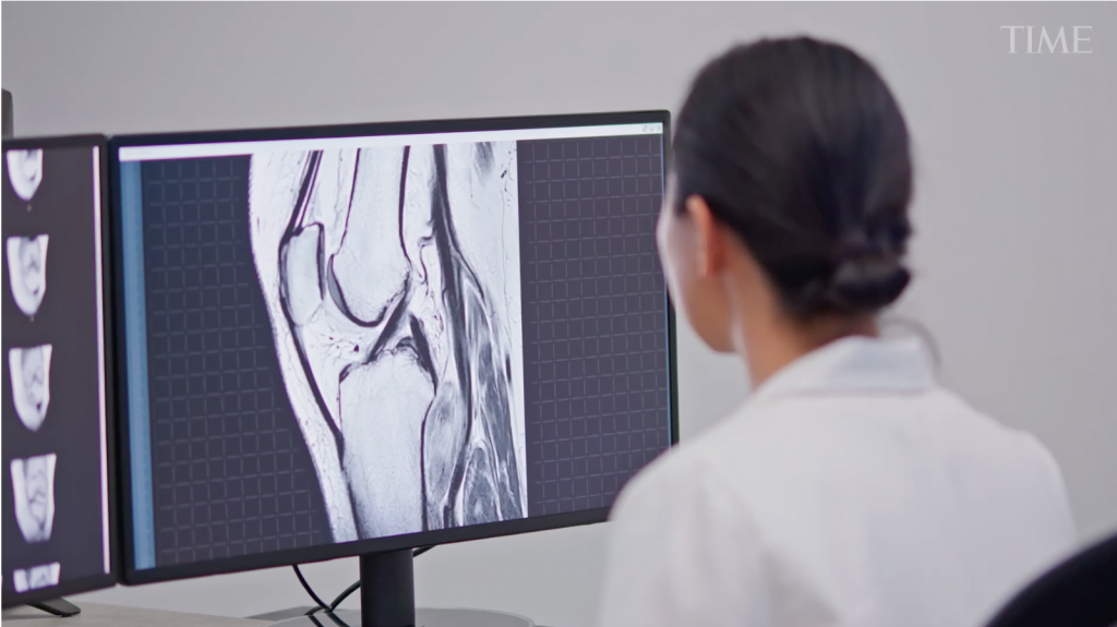
A medical professional depicted in a Time magazine video, reviews an x-ray which AI has already reviewed. (Source: Time.com)
Medical Imaging Takes Huge Leaps Forward with AI Reviews Drawing from Deep Data
The first x-ray is dated back to November 8, 1895, invented by Wilhelm Conrad Roentgen. From then on, the biggest problem in getting an x-ray was the heavy lead cover patients and techs would need to wear. The very idea of looking inside the human body was one that completely changed the world and in particular the world of medicine. Now that researchers have mixed AI with x-ray reviews based on past information, another paradigm shift in healthcare has occurred.
AI does a much better job working with and diagnosing conditions seen in x-rays. Adding AI to ultrasound equipment when searching for a tumor in a brain or a breast has shown a vastly improved rate of discovery. AI just reads the information better than humans do. Earlier treatment can be the difference between living and dying from cancer.
A story on time.com covers the giant leaps AI is making in the world of x-rays, MRI, CAT scans, and more. Writer Alice Park shares a wonderful quote from medical professional Dr. Keith Dreyer, chief data science officer and vice chairman of radiology at Mass General Brigham:
“The real change in health care that is going to happen from AI is that it will deliver a lot of solutions to patients themselves, or before they become patients, so that they can stay healthy,”
Imaging is also evolving from its initial focus—diagnosing medical conditions—to playing an integral part in treatment as well, especially in the area of cancer. Doctors are beginning to lean on imaging to help them monitor tumors and the spread of cancer cells so that they have a better, faster way of knowing if therapies are working. That new role for imaging will transform the types of treatments patients will receive, and vastly improve the information doctors get about how well they’re working so that they can ultimately make better choices about what treatment options they need.
Three of the major improvements doctors are seeing by using AI to help with imaging in the various types of scans inside bodies are these:
Detecting Problems Earlier
The first hurdle in making the most of what images can offer—whether they are X-rays, computerized tomography (CT) scans, magnetic resonance imaging (MRI), or ultrasounds—is to automate the reading of them as much as possible, which saves radiologists valuable time. Computer-aided algorithms have proven their worth in this area, as massive computing power has made it possible to train computers to distinguish abnormal from normal findings. Software specialists and radiologists have been teaming up for years to come up with these formulas; radiologists feed computer programs their findings on tens of thousands of normal and abnormal images, which teaches the computer to distinguish when images contain things that fall outside of normal parameters. The more images the computer has to compare and learn from, the better it becomes at fine-tuning the distinctions.
Tracking Patients More Closely
While computer-assisted triaging is the first step in integrating AI-based support in medicine, machine learning is also becoming a powerful way to monitor patients and track even the smallest changes in their conditions. This is especially critical in cancer, where the tedious task of determining whether someone’s tumor is growing, shrinking, or remaining the same is essential for making decisions about how well treatments are working.
Spotting Abnormalities
With enough data and images, these algorithms could even find aberrations for any condition that no human could detect, says Dreyer. His team is also working on developing an algorithm that measures certain biomarkers in the human body, whether anatomical or functional, so it can flag changes in those metrics that could suggest someone is likely to have a stroke, fracture, heart attack, or some other adverse event.
That’s the holy grail of imaging, says Dreyer, and while it’s a few years away, “those are the kinds of things that are going to be transformational in healthcare for AI.”
You can find more of Park’s article and information at the link below. The marriage of AI and x-ray tech is really one of the brighter tools that have been added to a physician’s ability to cure. And more importantly, the use of AI to find problems early.
“In the next five years, we will see functional imaging become part of care,” says Dr. Basak Dogan, associate professor of radiology at the University of Texas Southwestern Medical Center.
Dogan is testing two ways that imaging can be used to track functional changes in breast cancer patients. In one, using funding from the National Institutes of Health, she is imaging breast cancer patients after one cycle of chemotherapy to pick up slight changes in pressure around the tumor by injecting microbubbles of gas. Ultrasound measures changes in the pressure of these bubbles, which tend to accumulate around tumors; growing cancers have more blood vessels to support their expansion, compared to other tissues.
read more at time.com


Leave A Comment