Over 382,000 people are living with leukemia, or are in remission. Every day 170 Americans are diagnosed with leukemia and 67 lose the fight.
AI Sampling Cells Saves Substantial Time And Money
Perhaps the most astounding use of AI has been in the medical field. The changes and the insights AI is providing in nearly every department of a hospital, lab, or medical center continue to save more lives that once were impossible to save. Now comes news that an AI is diagnosing and helping to treat patients with leukemia.
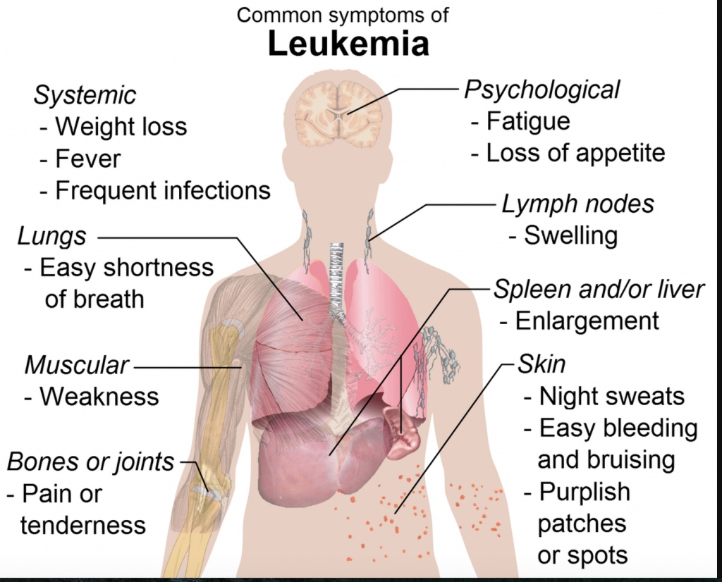
Leukemia is a broad term for cancers of the blood cells. The type of leukemia depends on the type of blood cell that becomes cancer and whether it grows quickly or slowly. Leukemia occurs most often in adults older than 55, but it is also the most common cancer in children younger than 15.
We found some new information about this particular AI algorithm at technologynetworks.com.
A team led by Prof. Dr. Peter Krawitz from the University of Bonn had already shown in 2020 that artificial intelligence can help with the diagnosis of such lymphomas and leukemias. The technology fully utilizes the potential of all measurement values and increases the speed as well as the objectivity of the analyses compared to established processes.
The method has now been further developed so that even smaller laboratories can benefit from this freely accessible machine learning method—an important step towards clinical practice. The study has now been published in the journal Patterns.
The article is heavily weighted with some serious medical terms and data that is a little deeper than some of the writing we share. For example:
Lymph nodes become swollen, patients experience weight loss and fatigue, as well as fevers and infections—typical symptoms of malignant B-cell lymphomas and related leukemias. If such cancer of the lymphatic system is suspected, the physician takes a blood or bone marrow sample and sends it to specialized laboratories. This is where flow cytometry comes in. Flow cytometry is a method in which the blood cells flow past measurement sensors at high speed. The properties of the cells can be detected depending on their shape, structure or coloring. Detection and accurate characterization of pathological cells is important when making a diagnosis.
The laboratories use “antibodies” that dock to the surface of the cells and are coupled to fluorescent dyes. Such markers can also be used to detect small differences between cancer cells and healthy blood cells. Flow cytometry generates large amounts of data. On average, more than 50,000 cells are measured per sample. These data are then typically analyzed on-screen by plotting the expression of the markers used against each other.
“But with 20 markers, the doctor would already have to compare about 150 two-dimensional images,” says Prof. Dr. Peter Krawitz of the Institute for Genomic Statistics and Bioinformatics at the University Hospital Bonn. “That’s why it’s usually too costly to thoroughly sift through the entire data set.”
All raw data and the complete software are open source and therefore freely accessible. In addition, res mechanica GmbH, which was involved in the study, has developed a web service that makes artificial intelligence usable even for users without bioinformatics expertise.
“With https://hema.to, we want to enable the exchange of anonymized flow cytometry data between laboratories and in this way create the conditions for even higher quality in diagnostics,” says Dr. Hannes Lüling of res mechanica.
If you are well versed in this field of medicine or if you are just interested because you know someone with this terrible disease, this article will give you lots of information and perhaps some hope for the future.
read more at technologynetworks.com


Leave A Comment